


 60 YEARS


4
March,
2026
I'm a Patient
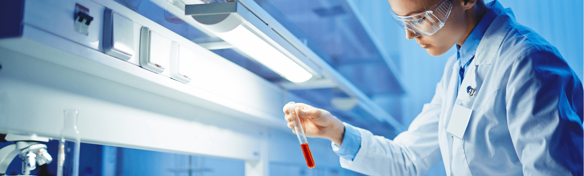
Your health is our priority. That’s why our goal is to provide caring, efficient, reliable and high-quality testing services to support you in effectively managing your health.
I'm a Healthcare Provider
LifeLabs is the largest provider of specialty laboratory testing services in Canada. Together with our partners, we provide national access to specialized clinical tests.
Our Tests
With over 70% of health care decisions based on diagnostic results, we know the importance of high quality tests. LifeLabs is committed to being a trusted health care partner and providing you with the information you need to make decisions.
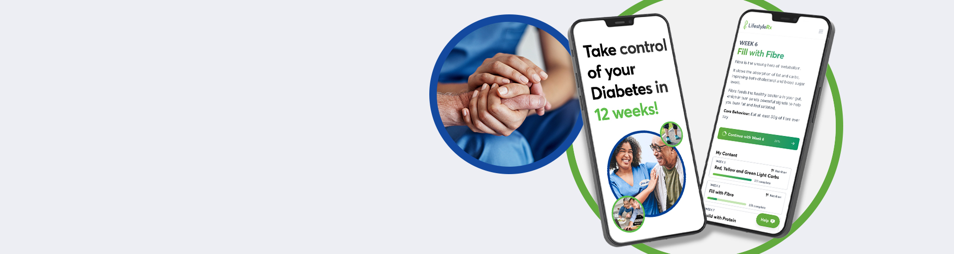
At-home Collection Kits
LifeLabs At-home Collection Kits let you order lab tests and collect your own samples at home - no help required! Order today and get the results you need to make informed decisions about your health.
Naturopathic Tests
Our Naturopathic division has a range of tests which help find trouble spots before the trouble really starts. From hormone levels, to food reactions, to identifying environmental toxins, we provide objective information so you can map your path to wellness.
Oncology
LifeLabs offers advanced cancer testing to help Canadians take a proactive approach to their health.
Our tests support early screening, identify inherited cancer risks, and provide key insights to guide treatment and monitor progress.
General Diagnostic & Genetic Tests
As Canada’s largest community lab, we offer thousands of diagnostic tests to ensure you have critical information about your health and to meet the diverse needs of the millions of patients who come to us each year for testing. Our genetic tests include a full suite of tests useful for planning your family and for diagnosing and monitoring cancer.

Empowering Healthier Canadians: How Caring in Action Makes a Differenc...

26
February,
2026
Empowering Healthier Canadians: Making Space for a Meaningful Differen...

18
February,
2026
Empowering Healthier Canadians: One Act of Kindness at a Time
At LifeLabs, we are proud Canadians, serving Canadians from coast to coast. In the “Empowering He...

12
February,
2026
Empowering Healthier Canadians: A Sisterhood Advancing Care

4
February,
2026
Empowering Healthier Canadians: How Caring Transforms the Patient Expe...
At LifeLabs, we are proud Canadians, serving Canadians from coast to coast. In the “Empowering Hea...

22
January,
2026